
Microscopic Anatomy and Organization of Skeletal Muscle Review Sheet 11 unveils the intricate architecture of skeletal muscle, providing a foundation for understanding its function and significance. Delving into the cellular and molecular components of muscle fibers, this review explores the fundamental principles that govern muscle contraction and adaptation.
The content of the second paragraph that provides descriptive and clear information about the topic
Introduction

Understanding the microscopic anatomy and organization of skeletal muscle is crucial for comprehending its function, mechanics, and pathology. Skeletal muscle, the primary tissue responsible for voluntary movement, exhibits a complex hierarchical organization, from the molecular level to the whole muscle.
Microscopic Anatomy of Skeletal Muscle: Microscopic Anatomy And Organization Of Skeletal Muscle Review Sheet 11
Components of a Skeletal Muscle Fiber, Microscopic anatomy and organization of skeletal muscle review sheet 11
A skeletal muscle fiber, the basic unit of muscle tissue, consists of several components:
- Myofilaments: Thin (actin) and thick (myosin) protein filaments that interact to generate force during muscle contraction.
- Sarcomeres: Repeating units within the myofilaments that represent the basic contractile unit of muscle.
- Myofibrils: Bundles of myofilaments arranged in a parallel fashion.
Organization of Muscle Components
Myofilaments are organized into sarcomeres, which are arranged in parallel to form myofibrils. Myofibrils are then bundled together to form muscle fibers. This hierarchical arrangement allows for efficient force generation and transmission.
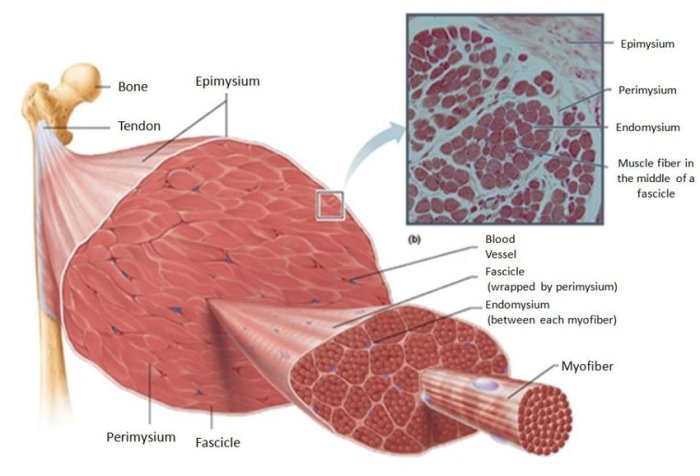
Connective Tissue Support
Skeletal muscle fibers are surrounded by connective tissue, which provides support and organization. The endomysium surrounds individual muscle fibers, the perimysium surrounds bundles of muscle fibers, and the epimysium surrounds the entire muscle.
Muscle Fiber Types

Skeletal muscle fibers are classified into different types based on their contractile properties and metabolic characteristics:
- Type I (Slow-twitch): Slow-contracting, fatigue-resistant, and aerobic-dominant.
- Type IIa (Fast-twitch oxidative-glycolytic): Fast-contracting, moderately fatigue-resistant, and capable of both aerobic and anaerobic metabolism.
- Type IIx (Fast-twitch glycolytic): Fast-contracting, fatigue-prone, and primarily anaerobic.
The distribution and function of each fiber type vary depending on the specific muscle’s function and demands.
Neuromuscular Junction

The neuromuscular junction is the site where motor neurons communicate with muscle fibers, initiating muscle contraction.
Structure
The neuromuscular junction consists of the motor neuron terminal, the synaptic cleft, and the muscle fiber membrane. The motor neuron releases neurotransmitters (e.g., acetylcholine) into the synaptic cleft, which bind to receptors on the muscle fiber membrane.
Neuromuscular Transmission
Binding of neurotransmitters triggers an action potential in the muscle fiber membrane, which spreads along the fiber’s surface and into the interior, causing calcium release from the sarcoplasmic reticulum. This calcium binds to troponin, initiating the sliding of myofilaments and muscle contraction.
Muscle Metabolism
Muscle contraction requires energy, which is primarily derived from the breakdown of glucose through anaerobic and aerobic pathways:
Anaerobic Metabolism
In short bursts of high-intensity activity, muscles rely on anaerobic metabolism, converting glucose to lactate without oxygen. This process produces ATP quickly but results in muscle fatigue.
Aerobic Metabolism
During sustained activity, muscles switch to aerobic metabolism, using oxygen to oxidize glucose and fatty acids for ATP production. This process produces more ATP but at a slower rate.
Muscle metabolism is regulated by hormones, neural signals, and the availability of oxygen and substrates.
Questions Often Asked
What are the key components of a skeletal muscle fiber?
Myofilaments (actin and myosin), sarcomeres, and myofibrils
How are muscle fibers classified based on their contractile properties?
Type I (slow-twitch), Type IIa (fast-twitch oxidative), Type IIx (fast-twitch glycolytic)
What is the role of the neuromuscular junction?
Transmits nerve impulses to muscle fibers, initiating muscle contraction
